Endonasal medial maxillectomy
Medial endonasal maxillectomy is the removal of the medial intersinonasal septum, which corresponds to the medial wall of the maxillary sinus in contact with the nasal cavity. This surgical procedure provides a wider, more direct view of the maxillary sinus cavity than a simple middle meatotomy. It is indicated almost exclusively in the management of benign tumors such as schneiderian papillomas, the most common of which is the inverted papilloma: 62% of cases, compared with 32% for exophytics and 6% for oncocytics.
Find out more about the endonasal medial maxillectomy procedure from Dr. Delagranda, ENT surgeon in La Roche sur Yon, France.
Who is concerned by endonasal medial maxillectomy?
Endonasal medial maxillectomy concerns adults with schneiderian papillomas, 62% of which are inverted papillomas.
It should be performed in cases of :
- Clinical signs associated with the presence of a papilloma: blocked nose, running down one side of the nostril and into the throat (nasal obstruction, anterior and posterior rhinorrhea), purulent discharge in the case of superinfection, headache (cephalalgia), diminished sense of smell (hyposmia), facial pain, unilateral nosebleed (epistaxis).
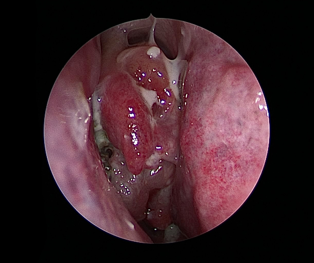
- Positive biopsy on a pink to red poly-lobed nasal mass that bleeds easily: patients are sometimes totally asymptomatic (4-23% of cases), and the tumour may have been discovered incidentally as part of another work-up (e.g. cerebral MRI).
Diagnosis is often delayed, averaging between 1 and 4 years after the appearance of the first signs, because patients are not bothered by the disease and are slow to seek help. Any persistent unilateral signs should prompt prompt consultation of an ENT specialist.

Intersinusonasal septum, inverted papilloma
Intersinusonasal septum
The intersinonasal septum is an irregular bony wall made up of several small bones (ethmoid, inferior turbinate, maxilla) covered with mucous membrane and perforated with orifices (obligatory or accessory) for communication between the maxillary sinus and the nasal cavity.
Inverted papilloma
Papillomas account for 0.5-4% of nasosinus tumours, affecting between 0.2 and 1.5 new cases per 100,000 inhabitants per year. Inverted papillomas are the most common, developing preferentially on the intersinonasal septum in men between the ages of 40 and 60 (5 men for 2 women). This benign tumor is locally aggressive and has a strong tendency to recur (5-60% according to studies), even over the long term (maximum in the first 3 years, but up to 10 years), but it also has the potential to transform into a malignant tumor or cancer of the squamous cell carcinoma type in 5 to 15% of cases. The HPV virus is associated with it in 40% of cases, especially in cancerous forms (serotypes 16 and 18). The bone opposite the foot of the inverted papilloma is often osteocondensed. CT (iso, homogeneous and microcalcifications in 20% of cases) and MRI (hypoT1 pdc+, cerebriform, iso-hypo T2) are recommended. A number of classifications exist, the most widely used of which is that of Krouse. Biopsy is mandatory to confirm the diagnosis, which is not always easy due to frequent associated benign polyps.
Exophytic papillomas never develop into cancer, and tend to develop on the medial wall of the nasal cavity.
Oncocytic papillomas have the same characteristics as inverted papillomas in terms of localization and malignant transformation, but develop into undifferentiated carcinomas.
Objectives of endonasal medial maxillectomy
- Eliminate pain.
- Improve the sensation of a blocked nose.
- Reduce anterior and posterior rhinorrhea and their consequences.
- Stop nosebleeds (epistaxis).
- Avoid future complications linked to the evolution of the benign tumor (deformations, infections).
- Remove benign or malignant tumors.
The different stages of the intervention
The surgical procedure
Under general anaesthetic in the operating theatre, the nasal cavities are cleaned with an additional anaesthetic, the ostium of the maxillary sinus is located, the dangerous anatomical landmarks are palpated in order to spare them, and a small bone forming part of the ethmoid in the intersinuso-nasal septum – the processus unciforme – is removed. With the unciform process removed, the new, larger opening includes the sinus ostium. This large opening allows the interior of the maxillary sinus to be inspected and cleaned, using angled optics (30° and 70°) and appropriate forceps and suction before continuing the procedure. The incision is then made in front of the inferior turbinate head, and the frontal process of the maxillary bone is motor-milled. The lacrimal duct is cut, and progress is made backwards, removing the entire inferior turbinate and papilloma. The very wide opening enables the effectiveness of the procedure to be checked using the optics. Healing foam is applied.
Post-surgery recovery period
In the case of outpatient surgery, the patient usually returns home the same day.
After hospitalization, you’ll need to rest at home for 7 days, and check that there’s no bleeding from the nose or throat.
If necessary, the surgeon will give you a 15-day medical leave.
Sport is not recommended for the first 15 days, and should be resumed gradually.
Pain is very moderate. It is controlled by Class I analgesics.
Post-operative care at home: nosewash with saline, analgesics, antibiotics if required by your doctor.
Scarring: no visible scar
Complications associated with endonasal medial maxillectomy
In addition to the risks inherent in any surgery involving general anaesthesia, endonasal medial maxillectomy presents rare complications:
- Nasal haemorrhage (epistaxis) after the procedure, which is very minor and rapidly subsides with nose-blowing and nose-washing.
- Periorbital hematoma.
- Air trapped in the eyelids (emphysema).
- Infection.
Frequently asked questions
Here is a selection of questions frequently asked by Dr Delagranda’s patients during consultations for endonasal medial maxillectomy in La Roche-sur-Yon.
Is surgery compulsory?
Yes, spontaneous evolution is slow but always negative, with a risk of transformation into cancer (5-15% of cases).
Is the effect long-lasting?
Yes.
Do I need regular check-ups afterwards?
Yes, because schneiderian papillomas frequently recur.
Is it necessary to be followed up for a long time afterwards?
Yes, because recurrences can occur up to 10 years later. One ENT consultation a year is recommended, along with an MRI, depending on the surgeon’s opinion and the technical difficulties of monitoring.
Is it painful?
Class I analgesics are generally sufficient.
Is postoperative care lengthy?
Yes. In particular, nosewashing must be continued for several weeks, as the exposed bone surface is quite large and generates substantial scabs in the nose.
Fees and coverage of the procedure
Medial endonasal maxillectomy is covered by the French health insurance system. Please contact your health insurance company to find out whether any extra fees will be covered.
Do you have a question? Need more information?
Dr Antoine Delagranda will be happy to answer any questions you may have about endonasal medial maxillectomy. Dr Delagranda is a specialist in ENT surgery at the Clinique Saint Charles in La Roche-sur-Yon in the Vendée.
ENT consultation for an endonasal medial maxillectomy in Vendée
Dr Antoine Delagranda will be happy to answer any questions you may have about endonasal medial maxillectomy. Dr Delagranda is a specialist in ENT surgery at the Clinique Saint Charles in La Roche-sur-Yon in the Vendée.

